Copanlisib in combination with nivolumab for microsatellite stable colorectal cancer: a phase 1/2 trial
Nature Communications. 2026;17:7114. DOI 10.1038/s41467-026-72985-6.
How to cite
AMA
Christenson ES, Wala JA, Parkinson R, Collins NB, Jakubowski C, Lin JR, et al. Copanlisib in combination with nivolumab for microsatellite stable colorectal cancer: a phase 1/2 trial. Nat Commun. 2026;17:7114. doi:10.1038/s41467-026-72985-6APA
Christenson, E. S., Wala, J. A., Parkinson, R., Collins, N. B., Jakubowski, C., Lin, J.-R., et al. (2026). Copanlisib in combination with nivolumab for microsatellite stable colorectal cancer: a phase 1/2 trial. Nature Communications, 17, 7114. https://doi.org/10.1038/s41467-026-72985-6BibTeX
@article{christenson2026copanlisib,
  title   = {Copanlisib in combination with nivolumab for microsatellite stable colorectal cancer: a phase 1/2 trial},
  author  = {Christenson, E. S. and Wala, J. A. and Parkinson, R. and Collins, N. B. and Jakubowski, C. and Lin, J.-R. and others},
  journal = {Nature Communications},
  volume  = {17},
  pages   = {7114},
  year    = {2026},
  doi     = {10.1038/s41467-026-72985-6}
}
In a phase 1/2 trial, the pan-PI3K inhibitor copanlisib was paired with the PD-1 inhibitor nivolumab in 39 heavily pretreated patients with metastatic microsatellite stable colorectal cancer — a setting that accounts for roughly 95% of metastatic disease and has historically not responded to single-agent checkpoint blockade. A molecularly defined subgroup achieved remarkable, durable clinical benefit: every partial response occurred in tumors carrying PI3K-pathway alterations, with progression-free survival spanning 24.2 to more than 33.7 months.
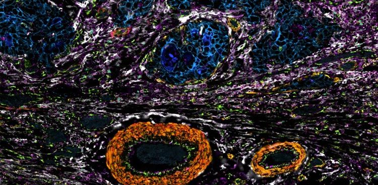
What set those responders apart was the question the study set out to answer — and answering it meant reading complex immune architecture from small metastatic core biopsies without exhausting the finite tissue a trial depends on. Investigators profiled 38 pre- and on-treatment core biopsies from 20 patients with RareCyte Orion 18-plex multiplex immunofluorescence, reading the tumor immune microenvironment at subcellular resolution from a single 5 μm tissue section.
Responders began treatment with more cytotoxic T cells already inside the tumor — a difference Orion could measure directly in scarce biopsy material — and the same imaging traced how those immune cells changed on treatment, and why recruitment into the tumor was not, on its own, enough.
Key findings
- On baseline biopsies, Orion resolved an immune signature that set responders apart. Across the 18 pre-treatment biopsies that met quality control, clinical responders carried significantly higher intratumoral CD3+CD8+ T-cell density and an elevated CD8+/FOXP3+ Treg ratio; and while PIK3CA-mutant tumors were broadly enriched for CD163+ macrophages, patients with a higher ratio of macrophage-to-tumor PD-L1 expression were significantly more likely to achieve a partial response.
- Paired biopsies showed treatment pulled T cells into the tumor, yet the tumor’s own signaling blunted the benefit. Intratumoral CD3+CD8+ T-cell infiltration rose in 100% of evaluable paired biopsies — 8 of 8 patients at 6 weeks (Cycle 2 Day 15) — corroborating within tissue the systemic expansion of activated CD8+ T cells that 34-plex mass cytometry (CyTOF) had registered in the blood. But Orion’s spatial view showed recruitment alone was not enough: localized, tumor-intrinsic ERK/MAPK signaling continued to drive resistance.
- Orion’s single-section read matched an orthogonal cyclic method on scarce biopsy tissue. Its quantitative T-cell infiltration measurements — from single-pass, 18-plex imaging of one 5 μm FFPE section at subcellular resolution — reached high analytical concordance when cross-validated against serial-section t-CyCIF on a RareCyte CyteFinder platform, so a single imaging round conserved finite core-biopsy material without giving up agreement.
Orion in the methods
“Orion multiplex immunofluorescence (IF) imaging uses specialized fluorophores with narrow excitation and emission spectra to provide for 18-plex IF in a single imaging run. We employed Orion multiplex IF to provide a basic characterization of the immune microenvironment of the full set of available specimens using a previously standardized antibody panel (Supplementary Table 1) and which have been previously validated. Unstained 5 µm thick FFPE slides were deparaffinized and stained with a fluorophore-conjugated IF antibodies. Stained slides were then imaged with our Orion instrument (RareCyte). Raw microscopy images were then stitched together with MCMICRO software.”
— Christenson et al., Nature Communications (2026), Methods, “Orion multiplex immunofluorescence”