High-plex immunofluorescence imaging and traditional histology of the same tissue section for discovering image-based biomarkers
Nature Cancer. 2023;4(7):1036-1052. DOI 10.1038/s43018-023-00576-1. PMID 37349501. PMCID PMC10368530.
How to cite
AMA
Lin JR, Chen YA, Campton D, Cooper J, Coy S, Yapp C, et al. High-plex immunofluorescence imaging and traditional histology of the same tissue section for discovering image-based biomarkers. Nat Cancer. 2023;4(7):1036-1052. doi:10.1038/s43018-023-00576-1APA
Lin, J. R., Chen, Y. A., Campton, D., Cooper, J., Coy, S., Yapp, C., et al. (2023). High-plex immunofluorescence imaging and traditional histology of the same tissue section for discovering image-based biomarkers. Nature Cancer, 4(7), 1036-1052. https://doi.org/10.1038/s43018-023-00576-1BibTeX
@article{lin2023highplex,
  title   = {High-plex immunofluorescence imaging and traditional histology of the same tissue section for discovering image-based biomarkers},
  author  = {Lin, J. R. and Chen, Y. A. and Campton, D. and Cooper, J. and Coy, S. and Yapp, C. and others},
  journal = {Nature Cancer},
  volume  = {4},
  number  = {7},
  pages   = {1036--1052},
  year    = {2023},
  doi     = {10.1038/s43018-023-00576-1},
  pmid    = {37349501}
}
Histopathology of hematoxylin and eosin (H&E)-stained tissue is still the main way cancer is diagnosed, while newer high-plex immunofluorescence adds spatially resolved, single-cell molecular data. The two usually come from different sections or different instruments, so aligning morphology and molecular signal cell by cell is hard.
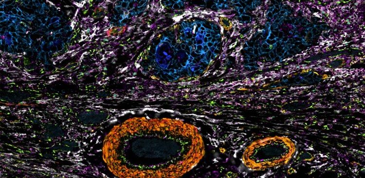
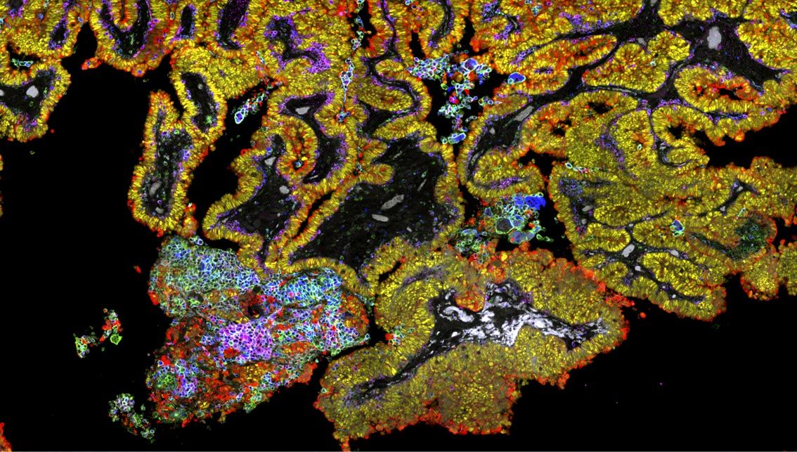
This paper introduces the Orion platform, which collects high-plex immunofluorescence and H&E from the same tissue section in a whole-slide format. Applied to 74 colorectal cancer resections in two cohorts, the paired images let pathologists and machine-learning models read complementary information and build spatial features predictive of progression-free survival, separating rapid from slow progression by 10- to 20-fold.
Key findings
- One-shot, whole-slide, 16- to 20-plex imaging on the same section as H&E. A commercial-grade Orion instrument used seven lasers (405 to 730 nm) and ArgoFluor-conjugated antibodies to acquire 16 to 18 antibody channels plus a nuclear stain and autofluorescence in a single round, followed by H&E staining and brightfield imaging of the same cells.
- Immunofluorescence and H&E carried complementary information. Molecular labels distinguished lymphocyte subsets that look identical in H&E, while H&E morphology recovered cell types the panel missed; 6.5 to 42% of nuclei (median 16%) could not be assigned an identity from immunofluorescence alone in the 40-specimen cohort.
- Spatial image features predicted colorectal cancer progression. Image-feature models built from Orion data recapitulated the Immunoscore, and searching combinations drawn from 13 immune markers produced a top model with a hazard ratio of 0.05 in cohort 1 (95% CI 0.02 to 0.10) that generalized to a second cohort (HR 0.17), a 10- to 20-fold discrimination of rapid versus slow progression across N = 74 participants.
Orion in the methods
“Whole slides were scanned on the Orion instrument using acquisition settings optimized for the specific antibody panels. Briefly, acquisition channel parameters were defined for each biomarker plus an additional channel dedicated to tissue autofluorescence and included excitation laser, emission CWL and exposure times.”
— Lin et al., Nature Cancer (2023), Methods, “One-shot antibody IF imaging with the Orion instrument”
Disclosure: RareCyte is listed as an author affiliation on the publication cited above.
Disclosure: RareCyte is named in the competing-interests statement of the publication cited above.
Why it matters for Orion users
If you are weighing Orion for tissue work, this is the study that defined what it does. The authors built a commercial-grade instrument that collects 16- to 20-plex immunofluorescence across a whole slide in a single staining round, then stains and images the same section with H&E, so every cell carries both its molecular identity and its morphology with no serial-section registration to reconcile. The single-round design is the point: the tissue is not carried through repeated stain-and-strip cycles, and the authors still read 16 to 18 antibody markers at once and recovered H&E their pathologists judged diagnostic grade from the same section. Here the payoff was concrete. Reading immune and tumor features in place across 74 colorectal cancer resections let the team recapitulate the Immunoscore and build spatial image-feature models predictive of progression-free survival. When your question is where cell types sit relative to the tumor boundary, and whether that arrangement forecasts outcome, you need the markers, the morphology, and the position measured on the same cells. That is the capability this paper established for Orion.