Novel Digital Pathology Algorithms to Explore the Role of Tumor Microenvironment in Next Generation Immuno-oncology Clinical Trials Utilizing 17-plex Fluorescent Immunohistochemistry
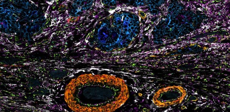
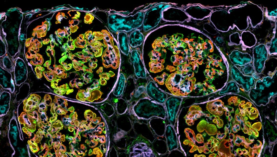
Linking the tumor microenvironment to response and resistance in immuno-oncology trials depends on turning spatial imaging into interpretable tissue structures; this AACR 2025 poster from Navigate BioPharma applies spatial cellular graph partitioning to single-step 17-plex Orion imaging and identifies 12 cellular microenvironments across five cancer types.
Presented by Navigate BioPharma Services.
Read or download to see:
- 12 cellular microenvironments were identified across five cancer types. Spatial cellular graph partitioning of segmented Orion imaging resolved neighborhoods ranging from single-lineage T-cell, B-cell and myeloid niches to tumor, tumor-infiltrated and checkpoint-enriched regions, mapped across non-small cell lung, breast, colorectal, prostate and melanoma samples (n=3 each).
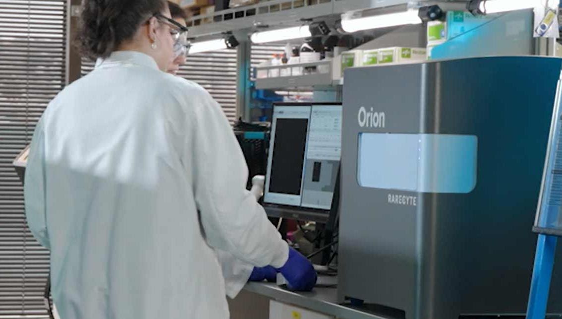
- Single-step Orion imaging feeds a graph-based neighborhood algorithm. The 17-plex assay combines 16 antibody markers characterizing T-cell, B-cell, myeloid, plasma-cell and tumor populations with a Hoechst nuclear counterstain, imaged in one Orion scan, then segmented and phenotyped in HALO and partitioned into cellular neighborhoods by spatial cellular graph partitioning (SCGP, described by Wu et al., 2024) before manual combination into the 12 microenvironments.
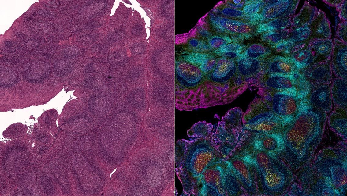
- Tonsil tissue anchored the approach as a proof of concept. In tonsil (n=3), spatial cellular graph partitioning recovered expected lymphoid structures, including follicle, germinal center, interstitium and crypt regions, indicating the method recapitulates known microenvironments; Navigate BioPharma reports the underlying 17-plex mFIHC assay was validated for clinical-trial use with automated IntelliPath FLX staining and Orion imaging.