Dr. Tanjina Kader on mapping spatial proteomics and chromosomal changes on the same tissue section
How Dr. Tanjina Kader pairs Orion™ single-round imaging with a modified DNA FISH protocol to read protein and chromosomal changes on one section of high-grade serous ovarian cancer.
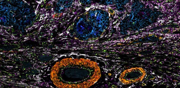
At the RareCyte booth at AACR 2026, Dr. Tanjina Kader of Harvard Medical School describes a method she developed to read both protein and chromosomal changes from a single tissue section. She images the section once on Orion, then reuses that same section for a modified DNA FISH protocol; because Orion images in a single round (“one shot,” in her words), the tissue adheres very well on the specimen. Kader studies how high-grade serous ovarian cancer progresses from normal cells, a cancer-evolution question that needs genetic information alongside the spatial protein view.
In this video:
- Dr. Kader studies how high-grade serous ovarian cancer progresses from normal cells, a question of cancer evolution that needs genetic as well as spatial information.
- She chose Orion because it images in a single round, which she describes as “one shot,” and which she says helps the tissue adhere very well on the specimen.
- The method she developed images a section on Orion, then runs a modified DNA FISH protocol on that same section, integrating protein and chromosomal changes at the same time.
- For research, the combined protein-plus-chromosomal readout supplies the genetic context that cancer-evolution studies require.
- For the clinic, Kader sees potential: because DNA FISH is used clinically, a validated risk-stratification biomarker could bring an Orion FISH protocol into clinical use.
Full transcript
Interviewer (RareCyte): We’re here at AACR at the RareCyte booth, and I’m here with Dr. Tanjina Kader from Harvard Medical School. First question for you: could you tell me a little bit about your research?
Dr. Tanjina Kader (Harvard Medical School): Yeah, sure, and thanks for having me. So I work on high-grade serous ovarian cancer, and I look into how high-grade serous ovarian cancer progresses from normal cells.
Interviewer (RareCyte): And how are you using the Orion platform?
Dr. Tanjina Kader (Harvard Medical School): So I actively work in the spatial proteomics field, and one of the platforms I use in particular is Orion, because Orion is one shot. It helps the tissue adhere very well on the tissue specimen. And I’ve also developed a new method where I use Orion, and after Orion I use the same section for a modified DNA FISH protocol, so it allows me to actually integrate protein and chromosomal changes together at the same time.
Interviewer (RareCyte): How do you see the Orion FISH protocol being used?
Dr. Tanjina Kader (Harvard Medical School): I think it has very much potential, definitely, to use in research settings, especially for someone like me who is interested in cancer evolution, because you do need some genetic information to understand cancer evolution. At the same time, it may have potential in the future to transition to the clinic, because in the clinic we already use the DNA FISH protocol. So it has high potential: if you have a biomarker for risk stratification of patients, for instance, you could actually use the Orion FISH protocol directly in the clinic.
Interviewer (RareCyte): Thank you so much.
Dr. Tanjina Kader (Harvard Medical School): Thank you.
Transcript reproduced from the recorded AACR 2026 booth interview (auto-generated captions), lightly edited only for speaker labels, obvious auto-caption artifacts, and the accuracy of proper names. The speaker’s name (Dr. Tanjina Kader) and the meeting name (AACR) were corrected from garbled captions; an unintelligible caption fragment was removed from one question. Punctuation was added during cleaning; the speakers’ words are otherwise as spoken.