Orion Spatial Proteomics Instrument
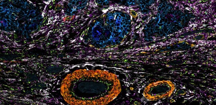
Orion images 20 channels per round across a whole slide at sub-micron resolution, giving spatial-biology researchers high-plex immunofluorescence, IHC, and same-section H&E from one automated instrument.
Orion runs high-speed, whole-slide imaging at sub-micron resolution and generates quantitative image analysis in parallel with the scan. You skip the separate analysis pass, since the data comes off the instrument ready to quantify. Integrated brightfield optics capture H&E on the same section as the immunofluorescence panel, and multi-round imaging takes the base 20-channel-per-round configuration to higher plex when a study calls for it.
OMERO Plus data management supports multi-user collaboration across 28 TB of onboard scan storage, and the whole system sits on a benchtop — sized for shared facilities and large cohort work.
Read or download to see:
- 20 channels per round at sub-micron, whole-slide resolution. High-speed scanning runs at 4 minutes per cm² per channel through a 20X 0.8 NA objective, across 402–808 nm excitation and a 450–874 nm emission range.
- Same-section H&E and quantitative analysis, built in. Integrated brightfield optics image H&E on the same section as the immunofluorescence panel, and quantitative image analysis runs in parallel with the scan, written to 16-bit OME-TIFF.
- Multi-round capability for higher-plex panels. The base configuration images 20 channels per round and extends through additional rounds; a 2048 × 2048 sCMOS camera at 6.5 µm pixels, 95% QE, and 1.0 electron read noise records every channel.
- Built for shared, cohort-scale labs. OMERO Plus data management, 28 TB of scan storage, 10 GB ethernet, and a two-slide stage run from a 21 × 22 × 23.5 in (53.4 × 56 × 60 cm) benchtop footprint on a Windows 10 Enterprise workstation.